Team
Our research team includes 3 board-certified radiologists with strong research expertise, along with 2 PhD students, 4 fellows and 10 residents actively involved in the unit's research activities. We place a strong emphasis on engaging young radiologists in research projects, whether as part of their Medical Doctorate (MD) or in pursuit of more advanced academic work.

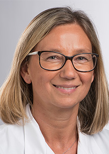
Pre Clarisse Dromain, MD, PhD
Médecin cheffe
Service de radiodiagnostic et radiologie interventionnelle
BH07
Bugnon 46
CH-1011 Lausanne
Contact e-mail
Fields of research
Oncologic Imaging – Biomarkers of tumor dynamics and imaging quantification
Her research focuses on the development of imaging-based biomarkers to quantify tumor behavior and treatment response, with particular emphasis on tumor growth kinetics and imaging heterogeneity. She explores how imaging—particularly advanced CT, MRI (including DWI and quantitative mapping), and PET/CT—can go beyond morphology to capture dynamic biological processes such as proliferation, necrosis, and vascular remodeling. A key axis of her work is the study of Tumor Growth Rate (TGR) as a continuous, quantitative biomarker of tumor aggressiveness, which we aim to validate across tumor types and integrate into prognostic models. She also investigates how radiomics and AI-driven extraction of spatial and temporal imaging features can improve response stratification, especially for treatments such as immunotherapy where conventional RECIST criteria fall short. Her group collaborates closely with biostatisticians and AI experts to develop interpretable models and assess their clinical relevance.
Translational Imaging Research in Digestive Oncology – NETs, peritoneal disease, and esophageal cancer
Her research program in digestive oncology focuses on translational imaging tools to better characterize and predict outcomes in complex tumor types, with an emphasis on neuroendocrine tumors (NETs), peritoneal carcinomatosis, and esophageal cancers. She develops and validates multiparametric imaging protocols (including hepatobiliary MRI, IVIM-DWI, and cine-MRI) to noninvasively assess tumor grade, vascularization, and growth patterns. In NETs, she investigates correlations between imaging phenotypes and histopathological or molecular markers, including somatostatin receptor expression and Ki-67 index, and she assesses tumor burden and heterogeneity using both conventional imaging and functional PET-based tracers. In peritoneal malignancy, she aims to create reproducible imaging-based criteria to guide surgical decisions and identify patients who will benefit from cytoreductive surgery. For esophageal cancer, she studies MR imaging surrogates of tumor regression grade post-therapy and work toward the integration of imaging and molecular data for response prediction. Her overarching goal is to bring quantitative imaging into the decision-making process through close integration with clinical trials and molecular pathology.

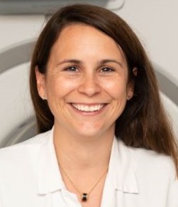
Dre Naïk Vietti Violi PD MER
Médecin associée
Service de radiodiagnostic et radiologie interventionnelle
BH07
Bugnon 46
CH-1011 Lausanne
Contact e-mail
Fields of research
Liver Imaging
Her research in liver imaging focuses on improving the early detection and treatment monitoring of hepatocellular carcinoma (HCC) and other liver diseases. Building on previous work, including ongoing collaborations with Mount Sinai Hospital (New York), she is leading a Swiss multi-year project funded by the SNF to evaluate new strategies for HCC screening. This includes comparing ultrasound, contrast-enhanced ultrasound, and abbreviated MRI (AMRI), as well as developing deep learning tools for automated detection including with cost-effective assumptions. She also pursues research on treatment response following loco-regional therapy, and on quantitative ultrasound and MRI techniques for evaluating liver function.
Advanced Imaging Methods
She is actively involved in advancing imaging methods for abdominal and oncology imaging applications, with a particular focus on artificial intelligence, radiomics, and low field MRI imaging development. Current projects include the use of radiomics to develop predictive models for characterization and prognosis for multiple diseases as chronic liver disease, HCC, pancreatitis and prostate cancer. Additionally, she is exploring the use of radiology in low-resource settings, with projects such as an ultra-portable ultrasound device for liver imaging and low-field MRI for prostate cancer detection. These innovations aim to transform radiology from qualitative image interpretation to a data-driven, personalized approach to diagnostics.


