Neuroradiology Unit
The Neuroradiology Unit is part of the Department of diagnostic and interventional radiology and provides medical services, pre/post graduate training, and conducts scientific research in diagnostic and interventional neuroradiology. Prof. Vincent Dunet, oversees the division's missions and activities. The medical staff is divided into two teams covering all the diagnostic (Prof Vincent Dunet, Prof Patric Hagmann, Dre Silvia Pistocchi, Dre Meriam Koob, Dr Gibran Manasseh) and interventional/therapeutic (Prof Guillaume Saliou, Dr Francesco Puccinelli, Dr Bruno Bartolini, Dr Steven Hajdu) aspects of brain and spine pathologies.
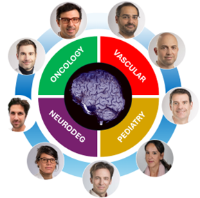
On the research side, all senior lecturers are involved into transversal collaborations with many other clinical departments inside the CHUV, with the Center for Biomedical Imaging (CIBM), and outside the CHUV with other national and international institute. Our research hence covers a wide range of pathologies over the lifespam from prenatal to elderly patients. This especially includes neurovascular diseases, brain and head and neck oncology, neurodegenerative and inflammatory diseases, traumatic brain injuries and fetal brain imaging.
Areas of investigation
Improving the diagnosis, management and prognosis of neurological diseases by utilizing advanced imaging and treatment techniques.
Neurovascular Imaging
Both diagnostic and interventional neuroradiologists of our team are involved in many local, national and international collaborations covering stroke diagnosis and treatment, as well as advanced treatment of cerebral aneurysm, arterio-venous fistula and arterio-venous malformations in the pediatric and adult population. These works are carried out in close collaboration with other departments of the CHUV, especially the Stroke Center and Department of Neurosurgery.
Neurodegenerative/Neuroinflammatory/TBI Imaging
Advanced brain imaging methods (automated morphometry, lesion segmentation, quantitative imaging, and brain connectivity) are developed to improve patients’ diagnostic and prognostic evaluation and management, in collaboration with the Department of Neurology, Leenards Memory Clinic, Siemens-CIBM-EPFL Laboratory and the Department of Nuclear Medicine and Molecular imaging.
Neurooncological and Head and Neck Imaging
Development and implementation of advanced brain multimodal imaging methods (automated detection, segmentation and characterization) to improve patients’ diagnosis and management, in collaboration with the Department of Oncology, the Department of Nuclear Medicine and Molecular imaging, the Department of Head and Neck Surgery, the Lundin Foundation, and the HES-SO.
Prenatal and Pediatric Neuroimaging
Development and implementation of advanced brain imaging methods (super resolution brain reconstruction, brain segmentation, brain connectivity) on high and low-field MRI scanner to improve detection and characterization of pediatric pathologies, and assess the normal and abnormal brain development, in collaboration with the Medical Imaging Analysis Laboratory (CIBM), the Departments of Gynecology and Obstetrics, Neonatalogy and Pediatrics.
Cross-cutting Themes
- Artificial intelligence and machine learning: Integrating these tools into image analysis for improved accuracy, efficiency, and decision support.
- Quantitative multimodal imaging: combining and evaluating respective performance and data-driven information provided by complementary modalities to provide personalized assessment,
- Personalized medicine: Tailoring imaging protocols and interpretations based on individual patient characteristics and risk factors, development of new therapeutic strategies and devices in the low field MRI environment.


Service de radiodiagnostic
et radiologie interventionnelle
Rue du Bugnon 46
CH-1011 Lausanne, Vaud, Suisse
+41 21 314 4444

Service de radiodiagnostic
et radiologie interventionnelle
Rue du Bugnon 46
CH-1011 Lausanne, Vaud, Suisse
+41 21 314 4444